14 Steps For A Supracondylar Amputation
The main goal of a supracondylar amputation is to obtain a stump that heals and without complications. The patient will thus be able to return to an almost normal life as soon as possible and with the best possible quality of life.
Supracondylar amputation is a surgical operation to cut a lower limb above the condyle. Between 50 and 65% of non-traumatic amputations of this type correspond to a complication of diabetes.
The very fact that a supracondylar amputation must be used implies the failure of any previous treatment of the problem, whatever it is. This requires a process of acceptance and the formulation of a clear goal for the patient: to protect their health and guarantee them the best possible quality of life.
The objective is thus to obtain a well healed, stable stump to which a prosthesis can be fitted as soon as possible. The ultimate goal is for the patient to return to normal life as soon as possible.
General principles
Generally, amputations fall into two categories: minor and major. The supracondylar amputation is major. Indeed, the limb to be amputated is of large size. However, whatever its degree or location, any amputation is a complex technical intervention that must be based on a few basic principles:
- Antibiotic treatment: this is prescribed as a preventive measure or to control an already existing infection
- Hemostasis, or control of bleeding, must be very rigorous: the appearance of a hematoma means the presence of necrosis or infection
- No tension at the anchoring points of the skin edges: to avoid this, it is necessary to handle the tissues with care
- Reasonable proportion between the bone section and the skin and musculotendinous length: this avoids tension and allows good bone coverage
- Traction of the nerve paths to prevent possible neuromas – benign nerve tumor – on the scar
- Ditto with articular cartilages and tendons
- Lack of bone chips in the wound or blunt edges
- Repeated washing of the surgical wound with physiological saline before proceeding with the suture
Indications and contraindications
Supracondylar amputation is performed when an infracondylar amputation has not healed properly. This is also the case with contracture of the calf muscles, which includes flexion in the knee joint.
Supracondylar amputation causes the loss of the knee joint. To avoid possible complications with the prosthesis that the patient will wear, it is important that the stump has an appropriate length.
This operation is not recommended in the presence of gangrene or an infection in the thigh.
Supracondylar amputation technique
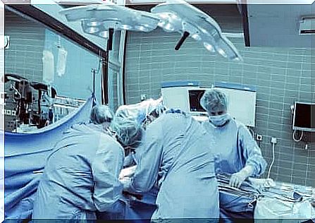
The steps to follow to perform a supdracondylar amputation are as follows:
- Place the patient in position supine dorsal
- Mark the incision as a “shark’s mouth”
- Cut the skin with a cold scalpel
- Section the hypodermis down to the aponeurosis. It is the membrane that covers the muscles and connects them to the bone. An electric scalpel is used for this. When severing in depth, enough tissue must be left for the stump
- Identification, ligation and respective section of the femoral vascular bundle and the sciatic nerve. It is necessary to carry out an infiltration of local anesthetic
- Surround the femur, encompassing its entire circumference
- Separate the tissues that are stuck to the femur. A periotome is used for this.
- Perform the adjustment of the Percy retractor for the amputation of the femur. This will allow enough soft tissue to remain to cover the bone stump
- Cut the bone using the Gigli saw. Take an angle of 90 ° between the two ends of the saw. During this time, the area should be washed continuously with physiological saline
- Then file the bony edges
- Apply bone wax -Horsley wax- to the section slice. We must apply the wax on the edge and stick it. Leftovers must be thrown away
- The stump is closed with a synthetic nonabsorbable monofilament Prolene® suture. The deepest muscle groups are sutured first to cover the bone surface. Then, the most superficial fascia is sutured
- The hypodermis is closed with an absorbable synthetic suture, better known as Vicryl®
- Finally, the skin is sutured with a silk suture, with mattress stitches